


광학,전자영상장비 공초점레이저주사현미경 A1Rsi
페이지 정보

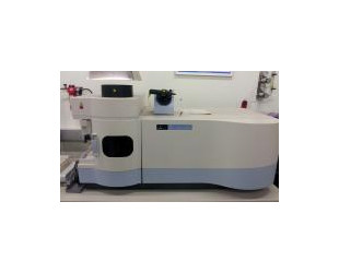
공초점레이저주사현미경 A1Rsi
Spectral Confocal Laser Scanning Microscope A1Rsi
시설장비활용번호Z-201907291106
시설장비등록번호NFEC-2012-03-155980
시설장비표준분류공초점현미경
- 장비활용서비스Biocon.Service
- 장비문의 031-888-9293
- 예약문의 032-888-8376
- 이용요금100,000원 ( 시간당 )
본문
장비정보
Information
- 제작사명(모델명)Nikon (A1Rsi)
- 구축일자2012년 02월 06일
- 사용용도시험
- 표준분류광학/전자영상장비 > 현미경 > 공초점현미경
- 설치장소인천광역시 연수구 송도과학로 85 포스코그린빌딩 1층 Lab1 F1층 lab1
- 장비설명고속과 고해상도의 두가지 scanner를 동시에 사용하여 기존의 confocal로는 관찰할 수 없었던 짧은 시간동안 일어나는 생물학적 변화를 고속촬영을 통해 관찰할 수 있으므로 매우 높은 quality의 이미지를 얻을 수 있을 뿐만 아니라 32 channel의 spectral detector를 이용하여 다양한 타겟에 대한 multiplexing이 가능함. 또한 live cell imaging에 필요한 장비들이 장착되어 있어 살아있는 상태의 세포를 장시간동안 관찰 분석할 수 있음
- 구성 및 성능1. Laser : BD 408nm Diode Laser Multi-line Ar Laser 488/514nm Solid State561nm LD 638nm
2. Detector
1) Standard fluorescence detector : 4 PMT
2) Diascopic detector : 1 PMT
3) Spectral detector: 32 channels
3. Scanning head
1) Standard image acquisition
Scanner : non-resonant scanner ×2
- Pixel size : max. 4096 × 4096 pixels
- Scanning speed : 4 fps (512 × 512 pixels)
2) High-speed image acquisition
Scanner : resonant scanner (X-axis) non-resonant scanner (Y-axis)
- Pixel size : max. 512 × 512 pixels
- Scanning speed : 30 fps (512 × 512 pixels) to 230 fps (512 × 64 pixels)
4. Monochrome cooled EMCCD
5. Cell incubation system
6. Eclipse Ti-E inverted fluorescence microscope
- Ultrahigh-speed imaging and photo activation
- FRET (Fluorescence Resonant Emerge Transfer)
- FRAP (Fluorescence Recovery after Photobleaching)
- FLIP (Fluorescence Loss in Photobleaching)
- Colocalization
- Three-dimensional time-lapse imaging
- Multipoint time-lapse imaging
- 사용/활용예
