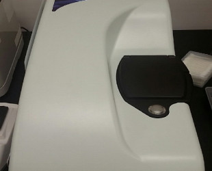
화합물전처리,분석장비 9.4T 자기공명영상장비
페이지 정보
9.4T 자기공명영상장비
9.4T MRI (Buruker BioSpec 94/20)
시설장비활용번호Z-202108314832
시설장비등록번호NFEC-2021-07-271775
시설장비표준분류핵자기공명분광기
- 장비활용서비스Cell to In-vivo 이미징 핵심연구지원센터
- 장비문의 032-899-6652
- 예약문의 032-899-6677
- 이용요금150,000원 ( 시간당 )
본문
장비정보
Information
- 제작사명(모델명)Bruker (Biospec 94/20 usr)
- 구축일자2008년 05월 05일
- 사용용도분석
- 표준분류화합물전처리/분석장비 > 분광분석장비 > 핵자기공명분광기
- 설치장소인천광역시 연수구 갯벌로 155 가천대학교 B1층 C004호
- 장비설명◯ MRI(Magnetic Resonace Imaging)는 원자핵(주로 수소)이 고유하게 방출하는 고주파 신호를 획득하여 컴퓨터를 통해 영상화 함.
◯ X-ray를 사용해 인체에 유해한 컴퓨터단층촬영 (computer tomography, CT)과 달리 MRI는 인체에 무해함.
◯ CT가 횡단면 주가되는 반면 MRI는 방향이 자유로움.
◯ 비침습적으로(non-invasively) 인체 내부의 해부학적 또는 병리학적 3차원 영상 정보를 세포 수준의 고해상도로 실시간 촬영이 가능함.
◯ 9.4T MRI는 실험동물의 해부학적 또는 병리학적 정보 영상(T1, T2, dMRI, fMRI)을 이용한 생체 분자이미징 활용 연구
◯ 다핵종 자기공명분광법(MRS) (1H, 31P, 13C, 23Na, 19F)을 통해 조직의 생화학적, 기능적인 영상정보 획득으로 대사물질의 정량화로 질병 진단 활용
- 구성 및 성능◯ Magnet
● Field strength: 9.4 T (BioSpec 94/20)
● Bore diameter: 20 cm
● Homogeneity (120 mm DSV): ± 10 ppm
● Magnet technology: Zero helium boil-off technology, nitrogen-free, Ultra Shielded and Refrigerated (USR) superconducting magnet for long hold-times, long maintenance intervals
◯ Gradients
● Gradient inner diameter: 114 mm
● Gradient strength: 660 mT/m with high power option
● Gradient insert with 1000 mT/m is available
● Slew rate: 3440 T/m/s (4570 T/m/s with high power option)
● Actively shielded gradient system
● Integrated shim coils for optimal field homogeneity
◯ ParaVision
ParaVision® 360: intuitive software package, for multi-dimensional MRI/MRS data acquisition, visualization, reconstruction, and analysis
● Over 100 validated and ready to use in vivo protocols and scan programs for mice and rats
● MRI sequence portfolio of more than 1,000 sequence variations, including wireless cardiac imaging using navigator based IntraGate methods with cartesian or radial readout, as well as short echo time imaging, such as UTE and ZTE
- 사용/활용예


